Brain Imaging, The Best Way To See Your Brain
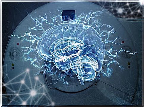
Technology has facilitated the development of multiple activities. Therefore, the health field has not been oblivious to these advances, helping to improve the diagnosis and treatment of patients. Without a doubt, one of the great advances in the field of medicine and psychology is the appearance of brain imaging.
With it, more precise information on the diagnosis can be obtained and, therefore, an intervention more adapted to each particular case can be planned.
In this approach, in addition, factors related to injury and its involvement in the brain are taken into account. But what are these tools? Next, we will show you some of the most used.
What are the advantages of brain imaging?
First of all, it is important to indicate that brain imaging helps to observe brain structure and function – it will do so depending on the technique used. It is important to indicate that its use is made both for the diagnosis and for the treatment of patients.
Using them you can identify where the damage occurs more quickly and accurately. With this, it is possible to analyze which would be the most appropriate treatment, depending on the location of the injury.
Likewise, the possible sequelae that the damage may have could be identified and thus know the best way to address them through multidisciplinary treatment.
During the rehabilitation processes, it is important to perform these types of exams. The results allow us to observe the new organization that occurs in the brain, how it compensated for the damage. In the end, you can see if there is any damage that could be identified at first or, if on the contrary, it has completely disappeared.
Computed tomography, structural brain imaging
In this technique an image is obtained through the use of X-rays. Multiple cuts are presented that allow the identification and location of basic structures, as well as some abnormalities.
This type of image is the most used when an emergency occurs. In essence, it serves to look at the general injuries that are most significant at the time of an accident.
One of the difficulties that can be observed with this type of brain imaging is that there is little differentiation between cortical and subcortical structures. Also, some of the brain structures cannot be seen in detail.
Magnetic resonance
This non-invasive technique uses radio frequency media and the magnetic field to image the brain.
It can be used both in the anatomical study of the brain and in the functional aspect, depending on what you are looking for.
Structural
Structural MRI, like computational tomography, allows you to see brain structures. In this case, the images are much more detailed, so that all parts of the brain can be seen, regardless of their size.
The results are shown from three perspectives, exposing multiple images of each of the slices. Therefore, it can help locate brain lesions. Its resolution is large, which makes it possible to identify small ones as well.
Another advantage that can be had with this type of brain imaging is being able to make quantitative measurements of brain structures .
These measures can be helpful in the diagnosis or treatment of diseases such as Alzheimer’s. The most notable thing is that the information is analyzed using several techniques, among which the following stand out:
- Voxels-based morphology analysis : in which the volume and concentration of gray matter and white matter are quantified and analyzed. As a result, it can be used to diagnose multiple disorders.
- Diffusion tensor imaging : in this, parameters are obtained that show the brain microstructures, especially those related to the white matter and its connections.
- Tractography : allows tracing the route of the main pathways of the white matter, knowing its route and possible damage.
Functional
It uses the same mechanism as the structural one, but its purpose is different. In this case, information about activation can be obtained when the brain is at rest or while carrying out an activity.
Its operation is based on the electrical activity of the brain. In these cases, areas that are performing a function produce more electrical activity compared to those that are not active.
Therefore, it is useful to know the affectation in the cerebral functionality. In addition, it provides information on some compensatory mechanisms in the development of the requested task. Similarly, in rehabilitation processes it has been used to identify the effects of a type of therapy.
PET and SPECT
Positron emission tomography (PET) and single photon emission tomography (SPECT) allow measurements of brain metabolism.
In them you can see regions that coincide functionally and structurally. This is due to the mild radiation that is included in the procedure.
Therefore, this type of brain imaging is useful in cases of head trauma : it can provide information on the affected region and the connections that could have been damaged. In this way, we can have a more precise knowledge about the consequences of the injury.
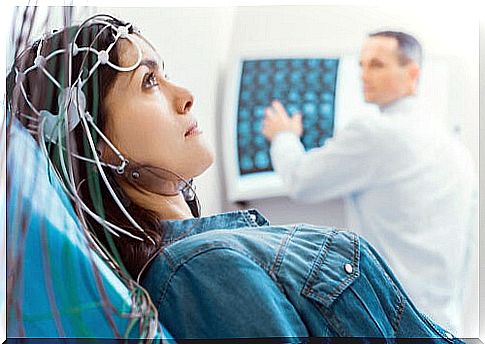
Electroencephalography
This technique uses the dendritic currents of neurons; that is, the energy emitted by them analyze their operation. Its information focuses on the registration of neurons present in the cerebral cortex. In this case, the results are presented in the form of waves, which adopt patterns that are related to some behaviors.
It is often widely used for the diagnosis of certain diseases, such as epilepsy or sleep apnea. Likewise, it is being used to measure and develop therapies such as neruofeedback.
In these cases, its use is useful to train neurons and thus regulate their functionality, mitigating the consequences of some disorders. In neuropsychological rehabilitation, it can be used as a treatment control, seeing changes in the waves of cortical connections.
Magnetoencephalography
This type of brain imaging allows us to know the magnetic fields of neurons during synapse. Therefore, brain activation can be directly observed during the development of an activity.
One of the advantages of this brain imaging technique is that it provides very precise temporal information. In other words, it limits the time it takes for our brain to activate when doing a task.
Currently, it is being used to investigate and understand how cognitive processes occur in the brain. Similarly, it is being used to investigate some of the premorbid symptoms of Alzheimer’s disease.
Why is brain imaging important?
With them, we can better understand what is happening in the brain. As we have said, they are very useful to individualize the intervention plan. Thus, having this variety of options, we can choose based on the demands of the patient and not so much on availability. However, it is no less true that there is a great but …
It is not a secret that its use is expensive for the health system. Therefore, magnetic resonance imaging is mainly used, since it is a tool that gives us structural and functional information. The positive part is that the results derived from research and medical practice with MRI have also been very satisfactory.